|
|
|
Main Menu
|
|
Sections
Meta
Talkback
Downloads
Information
|
|
|
|
|
|
Exposition: DNA structures from analyses of X-ray diffraction data
|
|
|
DNA structures from analyses of X-ray diffraction data
Authors: I.C. Baianu et al., contributors listed in the attached PDF file
Uploaded by:
bci1
|
- Comments:
- 2Mb, uploaded files
- Abstract:
- There are several alternate DNA structures from analyses of X-ray diffraction/scattering data. Thus, DNA exists in many possible conformations that include the A-DNA, B-DNA forms and the Z-DNA.(\cite{Ghosh}.) However, only B-DNA, and Z-DNA have been directly observed in functional (living) organisms. Which conformation DNA adopts depends on the hydration level, DNA sequence, the amount and direction of supercoiling, chemical modifications of the bases and also solution conditions, such as the concentration and type of metal ions and polyamines.
[{{cite journal |author=Basu H, Feuerstein B, Zarling D, Shafer R, Marton L |title=Recognition of Z-RNA and Z-DNA determinants by polyamines in solution: experimental and theoretical studies | journal=J Biomol Struct Dyn |volume=6 |issue=2 | pages=299–309 |year=1988 |pmid=2482766}}] Of these three DNA forms, the "B" form is most common under the conditions found in cells.[{{cite journal |author=Leslie AG, Arnott S, Chandrasekaran R, Ratliff RL |title=Polymorphism of DNA double helices |journal=J. Mol. Biol. |volume=143 |issue=1 |pages=49--72 |year=1980 |pmid=7441761 |doi=10.1016/0022-2836(80)90124-2}}] Note however that the `B-DNA form' is not a well-defined conformation but a family or fuzzy set of DNA-conformations that occur at the high hydration levels present in a wide variety of living cells.[{{cite journal |author=Baianu, I.C. |title=Structural Order and Partial Disorder in Biological systems|journal= Bull. Math. Biol. |volume= 42 |issue=4 |pages=464--468|year=1980|doi=10.1016/0022-2836(80)90124-2}}] Their corresponding (B-DNA) [[X-ray scattering techniques|X-ray diffraction and scattering patterns]] are characteristic of molecular [[Paracrystalline|paracrystals]] with a significant degree of disorder (>20%)[Hosemann R., Bagchi R.N., ''Direct analysis of diffraction by matter'', North-Holland Publs., Amsterdam--
New York, 1962][Baianu I.C., X-ray scattering by partially disordered membrane systems, ''Acta Cryst. A'', '''34''' (1978), 751--753.], and require a different approach from that of the standard analysis by [[Fourier transform]]s of [[Bessel function]]s [Bessel functions and diffraction by helical structures
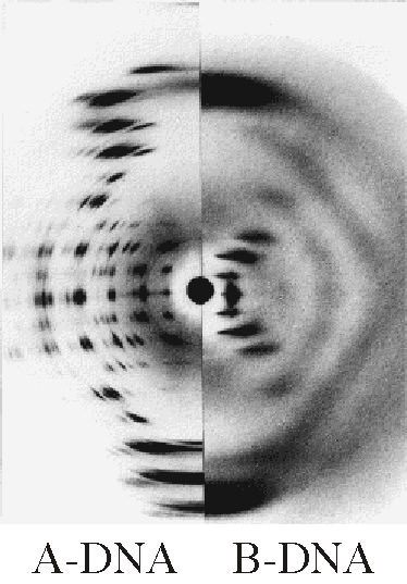
http://planetphysics.org/encyclopedia/BesselFunctionsAndTheirApplicationsToDiffractionByHelicalStructures.html]; the latter is routinely employed for the analysis of [http://www.genome.gov/Images/EdKit/picture_1943.gif A-DNA X-ray diffraction patterns] and Z-DNA X-ray diffraction patterns[X-Ray Diffraction Patterns of Double-Helical Deoxyribonucleic Acid (DNA) Crystals http://planetphysics.org/encyclopedia/BesselFunctionsApplicationsToDiffractionByHelicalStructures.html ] but is not suitable for the direct analysis of X-ray diffraction and scattering patterns of the B-DNA type. Thus, note the marked differences between the X-ray diffraction patterns of A- and B- DNA in the inserted image[[Image:ABDNAxrgpj.jpg|thumb| Comparison of the X-ray diffraction patterns of A- and B- DNA ]]([http://spdbv.vital-it.ch/TheMolecularLevel/BiochemViews/Nucleotides/Images/CMCC0904.jpg downloadable X-ray patterns of A-DNA and B-DNA] published by H. R. Wilson in 1958). Furthermore, the well-defined two double-helical forms of A- and Z- DNA can be shown to differ in their geometry and dimensions. The first published reports of A-DNA X-ray diffraction patterns-- and also B-DNA-- employed analyses based on [[Patterson function|Patterson transforms]] that provided only a limited amount of structural information for oriented fibers of DNA isolated from calf thymus[Franklin, R.E. and Gosling, R.G. received 6 March 1953. ''Acta Cryst''. (1953). '''6''', 673: The Structure of Sodium Thymonucleate Fibres I. The Influence of Water Content.; also ''Acta Cryst''. '''6''', 678: The Structure of Sodium Thymonucleate Fibres II. The Cylindrically Symmetrical Patterson Function.][{{cite journal| title=Molecular Configuration in Sodium Thymonucleate. Franklin R. and Gosling R.G| journal=Nature | volume= 171 | pages= 740–741 | year=1953 | url=http://www.nature.com/nature/dna50/franklingosling.pdf | pmid=13054694 | doi= 10.1038/171740a0| author=Franklin, Rosalind |format=PDF}}]. An alternate analysis was then proposed by Wilkins et al. in 1953 for B-DNA X-ray diffraction/scattering patterns of hydrated, bacterial oriented DNA fibers and trout sperm heads in terms of squares of Bessel functions[{{cite journal| title=Molecular Structure of Deoxypentose Nucleic Acids | author= Wilkins M.H.F., A.R. Stokes A.R. & Wilson, H.R. | journal=Nature | volume= 171 | pages= 738–740 | year=1953 | url=http://www.nature.com/nature/dna50/wilkins.pdf| pmid=13054693 | doi=10.1038/171738a0|format=PDF}}]
, and subsequently the latter X-ray diffraction patterns of DNA were re-analyzed by Crick and Watson with the help of [[molecular model]]s of a DNA double-helix[. The A-DNA form is a wider right-handed spiral, with a shallow, wide minor groove and a narrower, deeper major groove. The A form occurs under non-physiological conditions in partially dehydrated samples of DNA, while in the cell it may be produced in hybrid pairings of DNA and RNA strands, as well as in enzyme-DNA complexes.][{{cite journal |author=Wahl M, Sundaralingam M |title=Crystal structures of A-DNA duplexes | journal=Biopolymers |volume=44 |issue=1 | pages=45–63 |year=1997 |pmid=9097733 | doi = 10.1002/(SICI)1097-0282(1997)44:1 |doi_brokendate=2009-03-14}}][{{cite journal |author=Lu XJ, Shakked Z, Olson WK |title=A-form conformational motifs in ligand-bound DNA structures |journal=J. Mol. Biol. |volume=300 |issue=4 |pages=819-840 |year=2000 |pmid=10891271 |doi=10.1006/jmbi.2000.3690}}] Segments of DNA where the bases have been chemically modified by [[methylation]] may undergo a larger change in conformation and adopt the [[Z-DNA|Z form]]. Here, the strands turn about the helical axis in a [[left-handed]] spiral, the opposite of the more common B form.[{{cite journal |author=Rothenburg S, Koch-Nolte F, Haag F |title=DNA methylation and Z-DNA formation as mediators of quantitative differences in the expression of alleles | journal=Immunol Rev |volume=184 | pages=286–98 |year=2001|pmid=12086319 |doi=10.1034/j.1600-065x.2001.1840125.x}}] These unusual structures can be recognized by specific Z-DNA binding proteins and may be involved in the regulation of transcription.[{{cite journal |author=Oh D, Kim Y, Rich A |title=Z-DNA-binding proteins can act as potent effectors of gene expression in vivo |url=http://www.pubmedcentral.nih.gov/articlerender.fcgi?tool=pubmed&pubmedid=12486233 |journal=Proc. Natl. Acad. Sci. U.S.A. |volume=99 |issue=26 |pages=66-71 |year=2002 |pmid=12486233 | doi = 10.1073/pnas.262672699}}]
[[Image:Parallel telomere quadruple.png|thumb|left|300px|Structure of a DNA quadruplex formed by [[telomere]] repeats. The conformation of the DNA backbone diverges significantly from the typical helical structure.[Created from [http://ndbserver.rutgers.edu/atlas/xray/structures/U/ud0017/ud0017.html NDB UD0017]]]].\\
Hosemann R., Bagchi R.N., Direct analysis of diffraction by matter, North-Holland Publs., Amsterdam--New York, 1962.\\
Baianu I.C., X-ray scattering by partially disordered membrane systems, Acta Cryst. A, 34 (1978), 751--753. \\
Bessel functions and diffraction by helical structures: http://planetphysics.org/encyclopedia/BesselFunctionsAndTheirApplicationsToDiffractionByHelicalStructures.html ;
X-Ray Diffraction Patterns of Double-Helical Deoxyribonucleic Acid (DNA) Crystals: http://planetphysics.org/encyclopedia/BesselFunctionsApplicationsToDiffractionByHelicalStructures.html\\
Franklin, R.E. and Gosling, R.G. received 6 March 1953. Acta Cryst. (1953). 6, 673: The Structure of Sodium Thymonucleate Fibres I. The Influence of Water Content.; also Acta Cryst. 6, 678: The Structure of Sodium Thymonucleate Fibres II. The Cylindrically Symmetrical Patterson Function. \\
Franklin, Rosalind (1953). "Molecular Configuration in Sodium Thymonucleate. Franklin R. and Gosling R.G" (PDF). Nature 171: 740--741. doi:10.1038/171740a0. PMID 13054694. http://www.nature.com/nature/dna50/franklingosling.pdf. \\
Wilkins M.H.F., A.R. Stokes A.R. and Wilson, H.R. (1953). "Molecular Structure of Deoxypentose Nucleic Acids" (PDF). Nature 171: 738--740. doi:10.1038/171738a0. PMID 13054693. http://www.nature.com/nature/dna50/wilkins.pdf. \\
Wahl M, Sundaralingam M (1997). "Crystal structures of A-DNA duplexes". Biopolymers 44 (1): 45--63. doi:10.1002/(SICI)1097-0282(1997)44:1 (inactive 2009-03-14). PMID 9097733. \\
Lu XJ, Shakked Z, Olson WK (2000). "A-form conformational motifs in ligand-bound DNA structures". J. Mol. Biol. 300 (4): 819-840. doi:10.1006/jmbi.2000.3690. PMID 10891271. \\
Rothenburg S, Koch-Nolte F, Haag F (2001). "DNA methylation and Z-DNA formation as mediators of quantitative differences in the expression of alleles". Immunol Rev 184: 286-298. doi:10.1034/j.1600-065x.2001.1840125.x. PMID 12086319. \\
Oh D, Kim Y, Rich A (2002). "Z--DNA--binding proteins can act as potent effectors of gene expression in vivo". Proc. Natl. Acad. Sci. U.S.A. 99 (26): 16666-71. doi:10.1073/pnas.262672699. PMID 12486233. http://www.pubmedcentral.nih.gov/articlerender.fcgi?tool=pubmed&pubmedid=12486233.
- Rights:
-
Open access:
http://en.wikipedia.org/wiki/DNA
- Download:
-
- Links:
|
|
|
|
|
|
|
|
|
Pending Errata and Addenda
|
|
|
|
|
|
|
|
|
|
|
